-
 sudip.1583@gmail.com
-
 +91 86973 - 74363
1. Preparation: Remove jewelry, glasses, or metal objects.
2. Positioning: Stand or sit as instructed by the technician.
3. Placement: Bite on a tab or place the X-ray sensor in your mouth.
4. Exposure: Hold still while the X-ray is taken (painless).
5. Image Capture: Technician checks image quality.
6. Multiple Views: Repeat for different angles if needed.
7. Review: Dentist examines X-rays for diagnosis.
Bitewing X-rays: Check cavities and bone level.
Periapical X-rays: Show entire tooth.
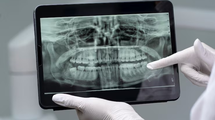
Panoramic X-rays: Full jaw view.
1. Detect hidden cavities or issues.
2. Assess bone health and tooth roots.
3. Plan treatments like braces or implants.
4. Monitor oral health over time.
1. Preparation (remove metal objects).
2. Positioning and placement of X-ray sensor.
3. Painless exposure (takes seconds).
4. Image review by dentist.
1. Minimal radiation exposure.
2. Lead aprons for protection (if needed).